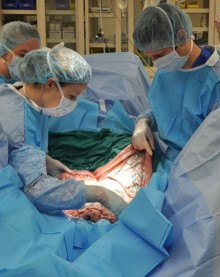
CALEC surgery, short for cultivated autologous limbal epithelial cells, signifies a groundbreaking development in eye injury treatment, particularly for those suffering from corneal damage that once seemed irreversible. Recently pioneered at Mass Eye and Ear, this innovative procedure utilizes the patient’s own limbal stem cells to regenerate the corneal surface, significantly enhancing vision and alleviating pain. During a clinical trial, 14 participants saw remarkable success, with CALEC surgery achieving over 90 percent effectiveness in restoring corneal integrity after 18 months. This approach not only paves the way for advanced stem cell therapy applications but also addresses the critical need for effective corneal surface restoration for patients with debilitating eye conditions. As researchers continue their efforts, the potential for CALEC surgery to transform the management of severe eye injuries is becoming increasingly evident.
Cultivated autologous limbal epithelial cell therapy, often referred to as CALEC surgery, represents a novel technique in the realm of ocular medicine aimed at healing corneal damage. By harnessing the regenerative abilities of limbal stem cells, this treatment offers a promising alternative for patients with severely injured corneas, particularly those who have exhausted conventional therapies. The innovative approach focuses on extracting healthy stem cells from one eye, cultivating them into a graft, and then transplanting this cellular tissue into the damaged eye. This methodology not only improves corneal surface restoration but also highlights the evolving field of stem cell therapy in ophthalmology. As clinical trials progress, the evidence supporting the efficacy of this technique continues to expand, illuminating a path toward improved visual health for many.
Introducing CALEC Surgery: A Breakthrough in Eye Repair
In a groundbreaking achievement, Ula Jurkunas conducted the first CALEC surgery at Mass Eye and Ear, marking an important milestone in ocular therapy. Cultivated Autologous Limbal Epithelial Cells (CALEC) surgery offers renewed hope for patients suffering from severe corneal damage—conditions once deemed impossible to treat. The procedure uses stem cells harvested from a healthy eye to restore the corneal surface, promoting healing and vision rehabilitation. This innovative approach not only treats the symptoms of corneal injuries but also addresses the underlying causes by regenerating limbal stem cells that maintain a healthy ocular surface.
The efficacy of the CALEC method has been recognized as more than just a temporary solution. With results showing over 90% success in restoring corneal surfaces, patients who once faced debilitating visual impairment are finding a path towards recovery. This surgery combines advanced stem cell therapy with pioneering surgical techniques, bringing together research and clinical practice in a way that has the potential to revolutionize eye injury treatment. By focusing on corneal surface restoration, the CALEC surgery thus sets a precedent within ophthalmology, highlighting the critical role of limbal stem cells in eye health.
The Role of Stem Cell Therapy in Eye Injury Treatment
Stem cell therapy has emerged as a pivotal element in the treatment of eye injuries, particularly in cases where traditional interventions fall short. The use of cultivated limbal epithelial cells in CALEC surgery exemplifies how regenerative medicine can play a vital role in eye care. By harnessing the regenerative properties of stem cells, this therapy not only aids in tissue restoration but also minimizes the need for more invasive surgical procedures, such as corneal transplants. Through clinical trials, researchers have documented significant improvements in patients’ visual acuity, thus affirming stem cell therapy’s role as a game-changer in ophthalmology.
Moreover, the increasing success rates observed in the CALEC trials highlight the importance of continued research and development in stem cell applications. As patients exhibited varying degrees of visual improvement, the data collected underscores the potential of stem cell therapy to address diverse ocular conditions. With a safety profile marked by minimal adverse events, the CALEC surgery represents a promising avenue not just for restoring vision, but also for enhancing the overall quality of life for individuals dealing with the aftermath of severe eye injuries.
Corneal Surface Restoration: Challenges and Innovations
Restoring the corneal surface is a complex challenge faced by ophthalmologists, especially for patients with limbal stem cell deficiency. Traditionally, severe injuries to the cornea have led to permanent visual impairment and ongoing pain due to the inability of the eye’s surface to heal itself. However, innovations such as CALEC surgery have opened new pathways for treatment. By utilizing stem cells from a healthy eye, clinicians like Ula Jurkunas are pioneering methods to regenerate vital ocular tissues that maintain transparency and structural integrity, crucial for proper vision.
Despite these advancements, challenges remain in terms of accessibility and the need for further research. Currently, the CALEC procedure is still experimental and not widely available, making it essential for ongoing studies to expand clinical trials. Efforts are being directed towards creating an allogeneic system that could source limbal stem cells from donor eyes, potentially allowing for broader treatment options for patients with bilateral injuries. The future of corneal surface restoration hinges on these innovations, promising to extend the life-changing benefits of stem cell therapy to a larger population in need.
Limbal Stem Cells: The Key to Eye Health
Limbal stem cells play a crucial role in maintaining the health and function of the cornea. Located at the junction of the cornea and sclera, these cells are essential for the regeneration of the corneal epithelium, ensuring that the eye maintains its protective barrier and contributes to overall visual clarity. When these stem cells are depleted due to trauma or disease, the cornea cannot heal on its own, leading to complex challenges in treatment. The significance of maintaining limbal stem cell populations has become central in eye injury treatment paradigms, especially in light of advancements like CALEC surgery.
Research at institutions such as Mass Eye and Ear has demonstrated the transformative potential of cultivating limbal stem cells for therapeutic purposes. The CALEC method exemplifies the transition from laboratory research to practical application, successfully translating scientific findings into clinical solutions. By focusing on the regeneration of limbal epithelial cells, this approach aims to address some of the most debilitating corneal diseases, driving advancements in personalized medicine and restoring the hope of vision in patients whose conditions were previously considered untreatable.
The Future of CALEC Surgery and Its Impact on Patients
As the CALEC surgery advances through clinical trials, the future holds significant promise for improving patient outcomes in ocular health. With a success rate of over 90% in restoring corneal surfaces, this innovative therapy marks a pivotal moment in ophthalmology, especially for those afflicted by severe corneal injuries. The collaboration between researchers, surgical teams, and manufacturing facilities exemplifies the integration of science and medicine, driving forward a treatment that could become the standard for eye injury repair. The long-term implications for patients are profound, potentially transforming the landscape of eye care.
In the coming years, researchers anticipate expanding studies to include larger populations and varying demographics, ensuring that CALEC surgery becomes widely accessible. The ongoing support from institutions like the National Eye Institute underlines the significance placed on this kind of research, emphasizing its relevance in developing advanced therapies. Moreover, as understanding of stem cell applications grows, the hope is that innovative procedures such as CALEC will not only enhance vision but also alleviate the suffering associated with chronic eye conditions, marking a new era in eye injury treatment.
Innovative Manufacturing Processes for Limbal Stem Cell Therapy
The development of CALEC surgery at Mass Eye and Ear has brought forth an innovative manufacturing process for cultivating limbal stem cells. This specialized technique involves taking a biopsy from a healthy eye, expediting the growth of these vital cells in a controlled environment, and preparing them for transplantation into the damaged eye. This meticulous process typically spans two to three weeks and ensures that the grafts meet stringent quality and safety standards required for patient use. The merging of biotechnology with cell therapy exemplifies the advanced methodologies being applied in modern medical treatments.
The innovation doesn’t stop at manufacturing; it extends to collaborative efforts between research institutions and hospitals. The partnership between Mass Eye and Ear and Dana-Farber Cancer Institute has been pivotal in perfecting the techniques needed to produce effective CALEC grafts. Through continuous research and development, this collaboration aims to overcome the current limitations of requiring a healthy eye for biopsy and move towards allogeneic sources for stem cells. This advancement would broaden the eligibility for future patients, exponentially increasing the potential range of treatments for corneal damage.
The Clinical Trial Journey: From Research to Reality
The journey of CALEC surgery from initial concept to clinical implementation underscores the rigorous process of medical research and trials. Since the first patient was treated in 2018, extensive studies have demonstrated the treatment’s promise in addressing corneal injuries. Participants in the trial showcased significant improvements in corneal health and visual acuity, with a noteworthy safety profile. The meticulous monitoring and evaluation throughout the trial phases elucidate the commitment to uphold patient care and safety while advancing medical science.
As this treatment proceeds towards potential FDA approval, the emphasis on large-scale, randomized trials remains pertinent. These studies are crucial for validating the long-term effectiveness and safety of CALEC surgery beyond the preliminary success observed. Since the trial is currently the first of its kind funded by the National Eye Institute, it holds substantial implications for the future of stem cell therapies in the field of ophthalmology. Achieving broader acceptance of CALEC surgery could transform care options available for individuals with acute corneal injuries.
Understanding Visual Acuity Changes Post-CALEC Surgery
A pivotal aspect of the success of CALEC surgery is the evaluation of its impact on visual acuity in patients post-treatment. In the initial study, improvements in vision were noted across all participants, confirming the procedure’s capability to restore not just the structural integrity of the cornea but also functional vision. For many individuals who had previously experienced ongoing pain and impaired vision, the newfound ability to see more clearly is not just a medical milestone but a profound personal transformation.
Regular assessments throughout the trial have shown that the rate of improvement in visual acuity can vary, yet the consensus remains that CALEC surgery offers substantial benefits to those suffering from corneal damage. Continuous monitoring will further clarify the long-term advantages of the procedure. As researchers seek to broaden the understanding of how stem cell therapy affects vision recovery, the findings may ultimately inform clinical best practices and patient management strategies following interventions for eye injuries.
The Collaborative Efforts Behind CALEC Research
The transformative research leading to CALEC surgery is the result of collaborative efforts involving multiple leading institutions, including Mass Eye and Ear, Dana-Farber, and Boston Children’s Hospital. This multidisciplinary approach combines expertise in ophthalmology, regenerative medicine, and cell manufacturing to address the urgent need for advanced treatments for corneal injuries. Researchers and clinicians have pooled their knowledge to develop a procedure that not only meets but exceeds current therapeutic standards, resulting in innovative solutions for previously untreatable conditions.
Such collaborative initiatives signify the essence of modern medical research, where shared objectives drive scientific inquiry and enhance treatment modalities. The joint efforts behind CALEC surgery also highlight the potential for future research in stem cell applications within ophthalmology and beyond. As ongoing partnerships evolve, the potential for broadening the impact of these advancements on patient care is promising, ensuring that cutting-edge therapies are accessible to those most in need of them.
Frequently Asked Questions
What is CALEC surgery and how is it related to stem cell therapy?
CALEC surgery, or cultivated autologous limbal epithelial cell surgery, involves harvesting limbal stem cells from a healthy eye, expanding them in the lab, and then transplanting them into a damaged eye. This innovative treatment, developed at Mass Eye and Ear, utilizes stem cell therapy to restore the cornea’s surface, providing new hope for patients with severe corneal injuries.
How effective is CALEC surgery for treating corneal injuries?
The CALEC surgery has shown promising results in clinical trials, with over 90% effectiveness in restoring the cornea’s surface for patients with previously untreatable corneal injuries. In trials, 50% of participants experienced complete corneal restoration within three months, which increased to 93% at 12 months.
Can CALEC surgery help with eye injuries caused by trauma or chemical burns?
Yes, CALEC surgery is specifically designed to treat eye injuries from trauma, chemical burns, infections, and other causes that lead to limbal stem cell deficiency. By restoring the corneal surface using stem cells, this surgery offers a solution for patients facing these challenging conditions.
What are limbal stem cells and why are they important in CALEC surgery?
Limbal stem cells are crucial for maintaining the cornea’s smooth surface and integrity. In CALEC surgery, these cells are harvested from a healthy eye to create a cellular tissue graft that is transplanted into a damaged eye, aiding in the restoration of vision and comfort for affected patients.
Where is CALEC surgery currently being performed, and is it widely available?
As of now, CALEC surgery remains in the experimental phase and is not widely available in hospitals. The procedure was first introduced at Mass Eye and Ear and is being studied through clinical trials, which are essential for obtaining federal approval.
What are the safety profiles associated with CALEC surgery?
The clinical trials for CALEC surgery have indicated a high safety profile, with no serious adverse events reported in either donor or recipient eyes. Minor complications, such as a bacterial infection, occurred in one participant but were resolved quickly, highlighting the surgery’s overall safety.
What advancements have been made in stem cell therapy for eye injuries at Mass Eye and Ear?
Mass Eye and Ear has made significant advancements in stem cell therapy, including the development of CALEC surgery to treat blinding corneal injuries. This innovative approach has been supported by extensive research, clinical trials, and collaborations with other institutions, showcasing the potential of stem cells in ocular health.
What is the future outlook for CALEC surgery and stem cell therapies in treating eye injuries?
The future of CALEC surgery looks promising, with ongoing research aimed at expanding its applications to patients with damage in both eyes. Researchers plan to conduct larger studies and seek FDA approval, with the hope of making this groundbreaking treatment accessible to a broader range of patients suffering from corneal injuries.
| Key Point | Details |
|---|---|
| Introduction of CALEC Surgery | Ula Jurkunas performed the first CALEC surgery at Mass Eye and Ear to treat cornea damage. |
| What is CALEC? | Cultivated Autologous Limbal Epithelial Cells (CALEC) is a stem cell treatment designed to restore corneal surfaces from blinding injuries. |
| Procedure Overview | The procedure involves taking stem cells from a healthy eye, expanding them into a graft, and then transplanting the graft into a damaged eye. |
| Clinical Trial Success | The treatment showed over 90% effectiveness in restoring the cornea in 14 patients over 18 months. |
| Patient Requirements | Patients must have only one eye involved to allow for a biopsy from the healthy eye. |
| Future Research | There are plans for larger studies and a goal to develop allogeneic manufacturing for broader patient eligibility. |
| Safety Profile | The procedure is considered safe, with minor adverse events and no serious complications reported. |
Summary
CALEC surgery represents a significant advancement in the treatment of corneal damage that was previously deemed untreatable. By utilizing stem cells from a healthy eye to restore the corneal surface, this innovative approach has shown promising results in a clinical trial, demonstrating a success rate of over 90%. With ongoing research and future trials, CALEC surgery has the potential to change the landscape of ophthalmic surgery and improve the quality of life for many patients suffering from severe corneal injuries.
